Quimica Clinica
View more documents from Paulina Alvarez.

![]()
CHRONIC OBSTRUCTIVE PULMONARY DISEASE
"HEAVES" - "BROKEN WIND" - "COPD"
- Introduction
 Chronic obstructive pulmonary disease (COPD) is an equine lung disease similar to human asthma. The clinical signs of COPD are caused by an allergic response to the particles in hay dust (see Figure 1). It is most often seen in older horses (greater than six years old) that are stabled during the winter months. COPD is rarely seen in warm, dry climates where horses are kept outside all year. Horses with COPD may exhibit clinical signs such as "heaving" to push air out of the lungs towards the end of exhalation, coughing, weight loss, and exercise intolerance. Wheezes may be heard towards the end of exhalation when listening to the airways with a stethoscope. A mucopurulent nasal discharge (composed of mucus and inflammatory cells) may be seen, especially after exercise. The abdominal muscles of COPD-afflicted horses may hypertrophy (enlarge) and form noticeable "heave lines." Heaves does not appear to be breed or gender related. There is evidence, however, that it may be hereditary.
- Etiology of COPD
Hay contains microorganisms such as bacteria and fungi as well as tiny particles of feed grains, plants, feces, dander, and pollen (see the photomicrograph of a clean hay sample in Figure 2). These tiny particles become aerosolized in hay dust and elicit an allergic response when they are inhaled by COPD horses. While it is believed that the hypersensitivity reaction seen in COPD horses is in response to many different allergens, the primary microorganisms involved in the etiology of heaves are Aspergillus fumigatus, Thermoactinomyces vulgaris, and Faenia rectivirgula. Aspergillus fumigatus is a mold that grows on dead and decaying matter such as poorly cured
  hay. It is thermophilic ("heat-loving") and can thrive in the high temperatures achieved in decomposing vegetation. A. fumigatus forms spores which become airborne and can be inhaled. These spores are antigenic (they are recognized as "foreign" by the immune system and provoke an immune response) and allergenic. Both Thermoactinomyces vulgaris and Faenia rectivirgula are bacteria which produce spores that become airborne and can be inhaled. All three of these species of microorganisms are numerous in moldy hay (Figure 3 is a photomicrograph of moldy hay).
- Pathogenesis of COPD
COPD is a disease that affects the air passages (trachea, bronchi, and bronchioles) through which air flows into the lungs (see Figure 4). The air passages are lined with layers of cells which constitute the epithelium. Below the epithelium is a layer of connective tissue called the submucosa. The epithelium and submucosa together are called the mucosa. Smooth muscle surrounds the bronchi and bronchioles all the way to the level of the alveoli (the air sacs in the lungs where gas exchange takes place). Contraction of the smooth muscle encircling the airways is known as bronchoconstriction or bronchospasm.
The airways are equipped with natural defense mechanisms to eliminate inhaled particles. These mechanisms include coughing, mucus secretion and removal, and bronchoconstriction. Chronic obstructive pulmonary disease is a delayed hypersensitivity reaction to inhaled allergens (materials that provoke allergic reactions). The natural defense mechanisms in the airways of COPD horses are hyperreactive and, therefore, they overreact when foreign particles are inhaled. Inflammation is also one of the defense mechanisms of the airways but in COPD inflammation occurs in excess and its purpose is not clear.
Inflammation
Four to six hours after a COPD horse is exposed to hay dust, the airways become inflamed and massive numbers of neutrophils accumulate within the air passages. Neutrophils are specialized white blood cells that kill bacteria. In COPD, it is still unclear what role the neutrophils play. It is known, however that the substances normally used by neutrophils to kill bacteria are capable of causing some of the changes in the airway epithelium observed in COPD horses.
 Each time a COPD horse is exposed to hay dust, its airways become acutely inflamed which causes the airways to become edematous (an abnormal accumulation of fluid in intercellular spaces). Repeated episodes of inflammation can cause the airway mucosal cells to proliferate. Both edema and proliferation of the mucosal cells thicken the airway walls and obstruct normal air flow during breathing (see Figure 5).
Mucus
 Airway mucus is produced in the trachea and bronchi by goblet cells in the epithelium and submucosal glands (see photomicrograph of goblet cells in Figure 6). Mucus lining the airways is viscous and sticky so that it entraps inhaled particles. The epithelium of the trachea and bronchi is covered with cilia (Figure 7 is a photomicrograph of ciliated epithelium). These tiny hairlike projections on the epithelial surface beat continuously and transport the overlying mucus layer up toward the larynx where the mucus can either be expelled (by coughing) or swallowed. The mucociliary system provides a means by which inhaled foreign particles can be cleared from the airways.
 Stimulation of the irritant receptors lying below the airway epithelium promotes mucus secretion so that more mucus is available to transport inhaled allergens out of the airways. Inflammation of the airways stimulates mucus secretion and causes proliferation of mucus producing cells. In COPD horses, excess mucus in the airways plugs the bronchioles (Figure 9 below is a photomicrograph of a mucous plug in the airways).
Bronchoconstriction
The smooth muscle that encircles the airways is controlled by the parasympathetic nervous  system. Inhaled irritants stimulate the parasympathetic nervous system to release acetylcholine (ACh). The binding of acetylcholine to receptors located on airway smooth muscle cells causes bronchoconstriction (bronchospasm) which prevents irritants from penetrating deeper into the lungs. When the mucosa is thickened by inflammation, even a little smooth muscle contraction can substantially narrow the airways and make breathing more difficult (this is illustrated in Figure 8). Air flow is also compromised by the increased production of mucus in response to inhaled allergens. Accumulated mucus and cellular debris in the airways further decreases the diameter of the air passages and increases the effort required to breathe. This increased work of breathing is evidenced
 by the abdominal push ("heaving") seen when COPD-afflicted horses try to force air out through the narrowed airways during exhalation.
Since the air passages of COPD-afflicted horses are obstructed (see Figure 9), oxygen cannot be efficiently delivered to the alveoli. This results in a low partial pressure of oxygen in the arterial blood of COPD horses. Less oxygen is available, therefore, for delivery to the tissues. Impairment of gas exchange in the lungs of COPD horses prevents these horses from performing well and results in exercise intolerance.
Cough
Coughing expels inhaled particles from the airways. Sensory nerve endings called irritant receptors lie below the airway epithelium. The irritant receptors are stimulated when inhaled particles or accumulated mucus secretions compress the airway epithelium and deform the underlying receptors. In COPD-afflicted horses, inflammation makes the cough reflex hyperreactive because the epithelium is damaged, irritant receptors become exposed, and the nerves become more sensitive to stimuli (much as an inflamed wound on your finger makes your finger more sensitive to touch). As a result of the hyperreactive cough reflex and mucus accumulation in the airways, COPD horses cough frequently (see Figure 10).
- Diagnosis
Veterinarians usually diagnose chronic obstructive pulmonary disease based on history and clinical signs. Since COPD is an allergic response to particles in hay dust, it should be determined how the horse is being housed and the type of feed it is receiving. Information supplied to the veterinarian by the owner or trainer about the onset and nature of clinical signs such as "heaving," coughing, or mucopurulent nasal discharge is also very useful. In addition, the veterinarian will want to know about any history of exercise intolerance.
The veterinarian will conduct a complete physical examination and pay particular attention to the lungs. Horses with COPD usually do not have a fever. Abnormal lung sounds, especially wheezing, become more obvious as the disease increases in severity. The veterinarian will look for evidence of a mucopurulent nasal discharge. If there is doubt about the diagnosis, the veterinarian may use endoscopy or bronchoalveolar lavage.
Endoscopy
Insertion of an endoscope through a nostril of the horse and into the trachea and bronchi allows the veterinarian to directly examine the air passages. The veterinarian will make note of any edema, hyperemia (the "redness" that occurs as more blood is shunted to areas of inflammation), and the presence and color of mucus accumulations. Figure 11 is a videoendoscopic photograph showing large amounts of mucous in the airways of a COPD-afflicted horse.
Bronchoalveolar Lavage
 Bronchoalveolar lavage is a process whereby a tube is passed through one nostril of the horse into the peripheral airways and then sterile saline is quickly injected and withdrawn from the air passages through the tube. This sample is then analysed microscopically for both the total number of cells present and the number and percentage of each cell type present (i.e. macrophages, lymphocytes, neutrophils, eosinophils, and mast cells). In normal horses, the predominant cells are macrophages and lymphocytes with neutrophils comprising less than five percent of all the
 cells present (see Figure 12). In horses with severe COPD, the percentage of neutrophils in bronchoalveolar lavage (BAL) fluid may be 50-70% (or more) of the total cell count (see Figure 13). However, horses with greater than 20% neutrophils will likely have impaired lung function and may have COPD.
Blood gas analysis can also be performed to assist in the diagnosis of chronic obstructive pulmonary disease. An arterial blood sample taken when the horse has just been exercised will have a lower partial pressure of oxygen than normal. Although horses usually become hypoxemic (low levels of oxygen in the blood) during exercise, the hypoxemia seen in COPD horses is more pronounced.
- Treatment of COPD
In equine COPD, inhalation of airborne allergens leads to airway inflammation which gives rise to bronchospasm. Treatment therefore involves prevention of exposure to allergens by environmental management, reduction of inflammation by use of corticosteroids, and relief of airway obstruction by use of bronchodilator drugs. Depending on the severity of the disease, use of the horse, and facilities available, one or all of these treatments may be used for a COPD-affected horse. There is no cure for COPD and, therefore, treatments need to be continued for life.
Environmental Management
The simplest way to treat a COPD horse is to change the environment so as to minimize exposure to hay dusts. This can easily be accomplished by putting the horse out to pasture. COPD-afflicted horses put out to pasture will go into clinical remission. If a horse must be stabled, then it is necessary to eliminate the use of straw for bedding and hay for feed. Even though the dust levels in the barn may seem insignificant, research has shown that the dust levels in the breathing zone (i.e. around the nose) of a horse eating hay can be as much as thirty to forty times higher than in the rest of the stall (see Figure 14). When a horse is eating a low dust feed such as pellets, the dust levels in the breathing zone are equivalent to those in the stall (see Figure 14). An effective management strategy for stabled COPD-afflicted horses, therefore, is to bed them on shavings and feed them a low dust diet. Feeds low in dust include complete pelleted feed, alfalfa cubes, and grass silage (haylage). Horses in adjacent stalls preferably should be kept in the same manner so as to prevent hay dusts from contaminating the stall of the COPD horse. However, if this is not possible, simply changing the management in one stall can dramatically improve lung function in a COPD-affected horse.
Hay should not be stored near the stall of a horse with COPD. Improving the ventilation in the barn can also help to minimize airborne particles. This may be accomplished by merely keeping the windows and doors open whenever possible or by using more sophisticated ventilation systems.
It is very important to realize that very short exposure of a COPD-susceptible horse to hay dusts can initiate inflammation and airway obstruction that can last for days. In a study by Fairbairn et al., COPD-susceptible horses were fed hay for seven hours and then put into a low dust environment. Three days later they still had inflamed airways . For this reason, COPD-affected horses should not simply be pastured during the day and then stabled and fed hay at night. This overnight exposure to dusts will be sufficient to maintain their airway obstruction.
Anti-Inflammatory Drug Therapy
In addition to changing the environment of a stabled COPD horse, it may be necessary to administer anti-inflammatory drugs. Corticosteroids are the drugs of choice for relieving inflammation of the airways. Corticosteroids can be administered by mouth, by injection, or by inhalation. When they are administered by mouth or by injection, therapy usually begins with a high dose and, as the horse improves, the dose is reduced to a maintenance level. Inhaled steroids offer the advantage of a high dose within the airways and minimal systemic side effects but a special mask is necessary for administration (see Figure 15). A chart outlining some of the more common corticosteroids used for COPD follows:
ANTI-INFLAMMATORY DRUGS:
| SUBSTANCE | MECHANISM | ADMINIS- TRATION | TREATMENT REGIMEN | POSSIBLE SIDE EFFECTS |
| Prednisolone | Corticosteroid | Oral or IM | Once daily; may administer every other day if clinical signs are controlled after a course of treatment | Few |
| Triamcinolone | Corticosteroid | IM | 1 dose every 3 months | Laminitis |
| Dexamethasone | Corticosteroid | Oral, IV or IM | Once daily for 2 days, then every other day | Laminitis |
| Beclomethasone diproprionate | Corticosteroid | Aerosol | Twice daily; after 2 weeks once daily | Few |
Bronchodilator Drug Therapy
Bronchodilators relax airway smooth muscle and relieve airway obstruction. In mildly affected horses, they may be the first line of therapy. They can also be safely combined with anti-inflammatory drugs for treatment of more severely affected horses. This combination is beneficial because anti-inflammatory drugs can reduce airway wall thickening but have no direct effect on the smooth muscle regulating the diameter of the airways. Bronchodilator drugs can be given orally, by injection, or by inhalation. Oral administration is the most convenient method but inhalation therapy is the most effective treatment for relief of airway obstruction. As with anti-inflammatory therapy, administration of bronchodilators by inhalation requires the use of a special mask (see Figure 15 above). Clinically useful bronchodilators drugs are outlined in the chart that follows:
BRONCHODILATORS:
| SUBSTANCE | MECHANISM | ADMINIS- TRATION | EFFECTS | DURATION OF ACTION | POSSIBLE SIDE EFFECTS |
| Clenbuterol (Ventipulmin syrup, Boehringer Ingelheim) (approved by FDA for use in horse, May '98) | b 2 adrenergic receptor agonist | Oral | Broncho- dilation; Stimulation of muco- ciliary escalator and mucus secretion | 12 hours | Tachy- cardia, sweating, excitation |
| Pirbuterol (Maxair) | b 2 adrenergic receptor agonist | Aerosol | Broncho- dilation; Stimulation of muco- ciliary escalator and mucus secretion | 1-2 hours | Minimal since adminis- tered by aerosol rather than systemically |
| Albuterol (Ventolin) | b 2 adrenergic receptor agonist | Aerosol | Broncho- dilation; Stimulation of muco- ciliary escalator and mucus secretion | 1-2 hours | Minimal since adminis- tered by aerosol rather than systemically |
| Ephedrine | Both a and b 2 receptors agonist | Oral | Broncho- dilation | Efficacy and duration never tested in clinical trials | Stimulation of central nervous system |
| Atropine | Nonspecific muscarinic antagonist | IV | Broncho- dilation | 4-6 hours | Gastrointestinal stasis and colic |
| Ipratropium bromide (Atrovent) | Quaternary ammonium nonspecific muscarinic receptor antagonist | Aerosol | Broncho- dilation | 4-6 hours | Minimal since poorly absorbed from airways into blood |
| Aminophyl- line (a derivative of theophylline) | Nonspecific inhibition of cAMP and cGMP phosphodi- esterases | Oral or IV | Broncho- dilation | Duration of effect never tested in clinical trials | Excitation, nervousness, increased heart rate at doses required for bronchodilation |
For information on the mechanism of action of bronchodilator drugs, click here.
This article was prepared by Rachele J. Baker under the direction of the faculty and staff of the Equine Pulmonary Laboratory.

|
| Urinalysis |
| Indications for performing this test: This test is often part of an initial data base for case work up of a clinically ill patient. It is a very useful indicator of renal function, and should be performed on any animal suspected to have renal disease or urinary tract pathology. A urinalysis should accompany a screening chemical panel for complete interpretation of the serum chemistries. Urinalysis is indicated in animals that have renal disease on their differentials list. |
Collection for analysis: There are several different methods of collection for urinalysis and each has its benefits and draw backs. Collection methods will often be dictated by the information that you are looking to gather.
Midstream: This collection method is often easiest for the animal but can be quite difficult for the collector. Collection is made into a container directly from the patient. This collection method will obviously contain contamination from the urethra and is therefore inadequate in the assessment of an upper urinary tract infection.
Manual Expression: This collection method is most often performed on small dogs and cats. It is sometimes difficult, and can result in trauma in the form of red blood cells in the urine. This method will also contain contamination from the lower urinary tract.
Catheterization: This test can be used on male dogs for the assessment of urethral patentcy and upper urinary tract infection. This method often results in iatrogenic presence of red blood cells in the urine.
Cystocentesis: This method requires penetration of the bladder through the body wall and can be accompanied by minimal bleeding. This is the best way to analyze the upper urinary tract for infection.
|
|
|
| Urethral catheterization being performed on a male dog. | Cystocentesis being performed on a male dog. |
The Test: The test is performed using a commercial dip stick to analyze most of the following parameters. Sedimentation is evaluated microscopically.
| Volume | Color | Turbidity | Odor |
| Specific Gravity | Sediment | pH | Glucose |
| Ketones | Bilirubin | Blood | Protein |
Volume
While it is difficult to evaluate volume based on a single sample, it is possible to do a 24 hour collection of urine to assess total urine production. Normal 24 urine production for dogs and cats is 20-44 ml/kg. An average sized saddle horse may produce between 5 and 15 L of urine in 24 hours. An increase in this volume is termed polyuria and may be due to physiological, pharmacological or pathological causes. Decreased urine volume is called oliguria, and occurs in dehydration, renal failure, or urinary blockages. No urine is called anuria, and is an emergency condition that may be due to renal failure, urinary blockage or ruptured bladder.
Keeping the urine in a calibrated container will
aid in determining the 24 hour volume.
 | Urine color will vary between species, but it is normally some shade of yellow depending on the concentration. Abnormal color changes in the urine could be due to drugs, increased urinary pigments or red blood cells. Red to reddish-brown could be due to either hematuria, hemoglobinuria, or myoglobinuria. Yellow-green to yellow-brown is associated with bilirubinuria. Occasionally, unusual colors may be caused by dyes associated with food or drugs. Pictured at left is a urine sample exhibiting hematuria. |
Urine is normally transparent in most animals, except for the horse. The horse has a thick viscous urine that is cloudy on examination. In small animals, turbidity suggests the presence of cells, casts, or crystals. Often refrigeration will propagate the sedimentation of crystals in the urine, producing a cloudy appearance. This is usually of no significance.
 | Here are two urine samples. The sample on the left is exhibiting turbidity. The sample on the right is a normal color and clarity for canine urine. |
Urine has a characteristic smell that varies slightly by species and concentration of the sample. A particularily foul odor may occur in the presence of bacteria. Thus, strong smelling urine is common in cases of cystitis. Ketonuria produces a very sweet smell as does glucosuria. Sweet smelling urine is commonly associated with acetonemia, pregnancy toxemia, and diabetes mellitus.
Do you smell that?
Specific gravity measures the concentrating ability of the kidney tubules. It is the ratio of the weight of urine to the weight of an equal volume of water. Normal values range from 1.001-1.060 in most of our domestic animals. If the kidneys are unable to concentrate urine the specific gravity will approach that of the glomerular filtrate, at 1.010. Hydration status will be reflected in urine specific gravity, therefore do not base profound observations of the renal concentrating ability on one specific gravity result.
 | This is a refractometer used to measure urine specific gravity. A small drop of the urine sample is placed under the glass slide on the top of the scope, and the measurement is made by looking through the eye piece to read the value indicated. |
| >1.030 | In dogs, a specific gravity this high indicates a normal concentrating ability, or perhaps dehydration. However, in cats, a specific gravity of this magnitude may accompany renal disease. To rule out renal disease in cats with concentrated urine, measure BUN and creatinine. If those values are in the normal range, you can likely rule out renal disease. |
| 1.013-1.030 | In dogs and cats without evidence of azotemia, this specific gravity is considered normal. If dehydration is suspected, values in this range may indicate abnormal concentrating ability, and further investigation in renal function should be made. |
| 1.008-1.012 | Urine specific gravity in this range is considered to be isosthenuric, meaning that is has not been concentrated in the tubules and is the same specific gravity as plasma. A water deprivation test may provide more information into the animal's concentrating ability. |
| <1.008 | Urine specific gravity in this range is termed, hyposthenuric, indicating the kidney's ability to dilute urine if necessary. In an animal with a need to diurese, this should be considered normal. However, in an animal that should be conserving water, this is highly indicative of renal disease. |
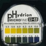
Urine pH will be affected by many things including the diet, handling of the sample, and acid-base balance of the animal. An alkaline pH is most indicative of an infectious process. Normal pH is between 6 and 8 for most animals depending on their diet.
| pH< 7.1 | pH in this range may be considered either acidic or normal. Carnivores who eat infrequently generally have a more acidic pH. If the pH drops below 6, then systemic acidosis should be considered. Other causes include: acidifying drugs, increased protein catabolism, and paradoxical aciduria associated with chloride and potassium depletion. |
| pH> 7.0 | Alkaline urine is produced postprandially and in urinary tract infections with Staphylococcus or Proteus spp. Alkaline urine is also common with renal tubular acidosis. |
In the normal animal there should not be glucose in the urine. If glucose is present, it is a classic response to hyperglycemia and should instigate an investigation into the possibility of diabetes in the patient. However, glucosuria can also occur without hyperglycemia. In these cases, it is due to a failure of the renal tubules to reabsorb the filtered glucose. A common associated finding with proximal renal tubular dysfunction is proteinuria. Proximal tubular dysfunction may be acquired or congenital as in some breeds of dogs.
In the normal animal there will be no ketones in the urine. An animal that is undergoing fat metabolism or is deficient in carbohydrates will have ketones in the urine. Slight ketonuria should be expected in malnourished animals. A ketonuria also frequently accompanies diabetes mellitus. Ketonuria will often precede detectable ketonemia.

Ketonuria can be detected with a simple U/A strip.
Bilirubinuria is caused by conjugated bilirubin, as unconjugated bilirubin does not filter through the glomerulus. Increased concentrations of conjugated bilirubin may be due to biliary obstruction, cholestasis, or increased production secondary to hemolysis.
Bilirubin levels in urine should be considered with urine specific gravity. A very concentrated urine with a trace of bilirubin carries much less significance than a dilute urine with some measure of bilirubin. Especially with concentrated urine, normal DOGS commonly have detectable bilirubin in their urine, but large amounts should not be present. Unlike dogs, bilirubinuria in CATS is always significant. However, bilirubinuria usually occurs in cats at about the same time that jaundice becomes apparent, so it is less valuable as a screening test.
Remember that bilirubinuria can occur with hemolytic disease, possibly via a "regurgitation" mechanism by which excessive unconjugated bilirubin presented to the liver is conjugated and then spilled into the systemic circulation as a result of saturated canalicular secretion.
There should not be any blood in the urine of a normal animal. Most test strips cannot differentiate between red blood cells, hemoglobin, or myoglobin, thus some care should be taken in interpretation. To determine which of these components is present, an examination of the serum should be made. If the serum is not red, it is unlikely to be due to hemoglobinemia. Myoglobinemia is rare in dogs and cats and should be accompanied by a clear serum and evidence of muscle trauma or disease. Hematuria is also evaluated in urine sedimentation microscopically and is reported as cells per high power field (or HPF). Remember that collection methods may also cause blood to appear in the urine. Other causes of hematuria include infection, neoplasia, or trauma.
Red blood cells present in a urine sample
Protein in the urine is a difficult assessment to make. It is a qualitative measurement rather than a quantitative measurement, and interpretation can vary between technicians. Protein should always be evaluated with knowledge of urine specific gravity. Concentrated dog and cat urine can contain small amounts of proteins. Proteinuria is always more significant in dilute urine. In significantly dilute urine, false negatives are possible. False positives for protein can occur in alkaline urine. In these cases, the sulfosalicylic acid test should be performed. The most accurate determination of proteinuria is the protein:creatinine ratio. Tubular concentration of urine increases the urinary protein and urinary creatinine concentrations equally so that the ratio remains constant whether the urine is concentrated or dilute. This ratio is normally less than 1. Proteinuria can be caused by inflammation, hemorrhage, or protein losing nephropathies.
Urine sedimentation may contain cells, casts and crystals and is examined microscopically after centrifugation of a urine sample. A very small amount of all of the above sediments is normal. Concern begins when any of these components is significantly elevated. There are many different crystals, cell types, and casts that may be found in the urine of animals, and it varies from species to species. Listed below are some common findings in the urine of small animals.
| Red & white blood cells | Less than 5 cells per high power field is considered normal. Any more than that in an animal other than a proestral bitch is considered abnormal. Causes of hematuria and/or pyuria include: trauma, uroliths, infection, neoplasia, parasites and coagulopathies. |
| Epithelial Cells | Epithelial cells are occasionally shed from the urethra, renal tubules, and bladder and are voided with the urine. Large clusters of epithelial cells are indicative of a transitional cell carcinoma. |
| Bacteria | The normal flora of the lower urinary tract may be shed with a voided sample. If urinary tract infection is suspected, a more sterile collection procedure should be used. Bacteria are not reliably seen until numbers reach 100,000/ml. Even then, it is difficult to correlate to an infection. Culturing the urine is the best method for establishing whether or not an infection is present. Common infectious agents of cystitis include: E. coli, staphylococci, streptococci, and Proteus spp. |
| Casts | Casts represent the normal turnover of tubular epithelial cells and are considered normal. However, large numbers of casts of either granular or hyaline types are considered abnormal. Increased granular casts are indicative of renal tubular cell injury due to many different causes. Increased hyaline casts are most often the result of glomerular proteinuria. |
| Crystals | Crystals may be considered normal or abnormal depending on the type and the species involved. In small animals, calcium oxalate dihydrate crystals and hippurate crystals suggest ethylene glycol toxicity. |
|
|
|
| Pictured above are Calcium Oxalate crystals. | These are also Calcium Oxalate crystals in a slightly different configuration. |
|
|
|
| Casts may appear in many different shapes and forms. | These two pictures (above right and left) are both of tubular casts. |
|
|
| Struvite crystals |
For more information . . .
Veterinary Laboratory Medicine. Duncan, Prasse, and Mahaffey page 162-174.
Veterinary Laboratory Medicine, Interpretation & Diagnosis. Meyer & Harvey, pages 221-232.
Manual of Small Animal Internal Medicine. Nelson and Couto, page 311.
Small Animal Medical Diagnosis. Lorenz and Cornelius, pages 590-599.

 embed-code