


leucemia linfocitica aguda en rufo.
Hace 15 años
|
| Urinalysis |
| Indications for performing this test: This test is often part of an initial data base for case work up of a clinically ill patient. It is a very useful indicator of renal function, and should be performed on any animal suspected to have renal disease or urinary tract pathology. A urinalysis should accompany a screening chemical panel for complete interpretation of the serum chemistries. Urinalysis is indicated in animals that have renal disease on their differentials list. |
Collection for analysis: There are several different methods of collection for urinalysis and each has its benefits and draw backs. Collection methods will often be dictated by the information that you are looking to gather.
Midstream: This collection method is often easiest for the animal but can be quite difficult for the collector. Collection is made into a container directly from the patient. This collection method will obviously contain contamination from the urethra and is therefore inadequate in the assessment of an upper urinary tract infection.
Manual Expression: This collection method is most often performed on small dogs and cats. It is sometimes difficult, and can result in trauma in the form of red blood cells in the urine. This method will also contain contamination from the lower urinary tract.
Catheterization: This test can be used on male dogs for the assessment of urethral patentcy and upper urinary tract infection. This method often results in iatrogenic presence of red blood cells in the urine.
Cystocentesis: This method requires penetration of the bladder through the body wall and can be accompanied by minimal bleeding. This is the best way to analyze the upper urinary tract for infection.
|
|
|
| Urethral catheterization being performed on a male dog. | Cystocentesis being performed on a male dog. |
The Test: The test is performed using a commercial dip stick to analyze most of the following parameters. Sedimentation is evaluated microscopically.
| Volume | Color | Turbidity | Odor |
| Specific Gravity | Sediment | pH | Glucose |
| Ketones | Bilirubin | Blood | Protein |
Volume
While it is difficult to evaluate volume based on a single sample, it is possible to do a 24 hour collection of urine to assess total urine production. Normal 24 urine production for dogs and cats is 20-44 ml/kg. An average sized saddle horse may produce between 5 and 15 L of urine in 24 hours. An increase in this volume is termed polyuria and may be due to physiological, pharmacological or pathological causes. Decreased urine volume is called oliguria, and occurs in dehydration, renal failure, or urinary blockages. No urine is called anuria, and is an emergency condition that may be due to renal failure, urinary blockage or ruptured bladder.
Keeping the urine in a calibrated container will
aid in determining the 24 hour volume.
 | Urine color will vary between species, but it is normally some shade of yellow depending on the concentration. Abnormal color changes in the urine could be due to drugs, increased urinary pigments or red blood cells. Red to reddish-brown could be due to either hematuria, hemoglobinuria, or myoglobinuria. Yellow-green to yellow-brown is associated with bilirubinuria. Occasionally, unusual colors may be caused by dyes associated with food or drugs. Pictured at left is a urine sample exhibiting hematuria. |
Urine is normally transparent in most animals, except for the horse. The horse has a thick viscous urine that is cloudy on examination. In small animals, turbidity suggests the presence of cells, casts, or crystals. Often refrigeration will propagate the sedimentation of crystals in the urine, producing a cloudy appearance. This is usually of no significance.
 | Here are two urine samples. The sample on the left is exhibiting turbidity. The sample on the right is a normal color and clarity for canine urine. |
Urine has a characteristic smell that varies slightly by species and concentration of the sample. A particularily foul odor may occur in the presence of bacteria. Thus, strong smelling urine is common in cases of cystitis. Ketonuria produces a very sweet smell as does glucosuria. Sweet smelling urine is commonly associated with acetonemia, pregnancy toxemia, and diabetes mellitus.
Do you smell that?
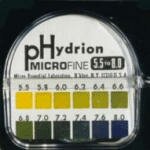
Specific gravity measures the concentrating ability of the kidney tubules. It is the ratio of the weight of urine to the weight of an equal volume of water. Normal values range from 1.001-1.060 in most of our domestic animals. If the kidneys are unable to concentrate urine the specific gravity will approach that of the glomerular filtrate, at 1.010. Hydration status will be reflected in urine specific gravity, therefore do not base profound observations of the renal concentrating ability on one specific gravity result.
 | This is a refractometer used to measure urine specific gravity. A small drop of the urine sample is placed under the glass slide on the top of the scope, and the measurement is made by looking through the eye piece to read the value indicated. |
| >1.030 | In dogs, a specific gravity this high indicates a normal concentrating ability, or perhaps dehydration. However, in cats, a specific gravity of this magnitude may accompany renal disease. To rule out renal disease in cats with concentrated urine, measure BUN and creatinine. If those values are in the normal range, you can likely rule out renal disease. |
| 1.013-1.030 | In dogs and cats without evidence of azotemia, this specific gravity is considered normal. If dehydration is suspected, values in this range may indicate abnormal concentrating ability, and further investigation in renal function should be made. |
| 1.008-1.012 | Urine specific gravity in this range is considered to be isosthenuric, meaning that is has not been concentrated in the tubules and is the same specific gravity as plasma. A water deprivation test may provide more information into the animal's concentrating ability. |
| <1.008 | Urine specific gravity in this range is termed, hyposthenuric, indicating the kidney's ability to dilute urine if necessary. In an animal with a need to diurese, this should be considered normal. However, in an animal that should be conserving water, this is highly indicative of renal disease. |
Urine pH will be affected by many things including the diet, handling of the sample, and acid-base balance of the animal. An alkaline pH is most indicative of an infectious process. Normal pH is between 6 and 8 for most animals depending on their diet.
| pH< 7.1 | pH in this range may be considered either acidic or normal. Carnivores who eat infrequently generally have a more acidic pH. If the pH drops below 6, then systemic acidosis should be considered. Other causes include: acidifying drugs, increased protein catabolism, and paradoxical aciduria associated with chloride and potassium depletion. |
| pH> 7.0 | Alkaline urine is produced postprandially and in urinary tract infections with Staphylococcus or Proteus spp. Alkaline urine is also common with renal tubular acidosis. |
In the normal animal there should not be glucose in the urine. If glucose is present, it is a classic response to hyperglycemia and should instigate an investigation into the possibility of diabetes in the patient. However, glucosuria can also occur without hyperglycemia. In these cases, it is due to a failure of the renal tubules to reabsorb the filtered glucose. A common associated finding with proximal renal tubular dysfunction is proteinuria. Proximal tubular dysfunction may be acquired or congenital as in some breeds of dogs.
In the normal animal there will be no ketones in the urine. An animal that is undergoing fat metabolism or is deficient in carbohydrates will have ketones in the urine. Slight ketonuria should be expected in malnourished animals. A ketonuria also frequently accompanies diabetes mellitus. Ketonuria will often precede detectable ketonemia.

Ketonuria can be detected with a simple U/A strip.
Bilirubinuria is caused by conjugated bilirubin, as unconjugated bilirubin does not filter through the glomerulus. Increased concentrations of conjugated bilirubin may be due to biliary obstruction, cholestasis, or increased production secondary to hemolysis.
Bilirubin levels in urine should be considered with urine specific gravity. A very concentrated urine with a trace of bilirubin carries much less significance than a dilute urine with some measure of bilirubin. Especially with concentrated urine, normal DOGS commonly have detectable bilirubin in their urine, but large amounts should not be present. Unlike dogs, bilirubinuria in CATS is always significant. However, bilirubinuria usually occurs in cats at about the same time that jaundice becomes apparent, so it is less valuable as a screening test.
Remember that bilirubinuria can occur with hemolytic disease, possibly via a "regurgitation" mechanism by which excessive unconjugated bilirubin presented to the liver is conjugated and then spilled into the systemic circulation as a result of saturated canalicular secretion.
There should not be any blood in the urine of a normal animal. Most test strips cannot differentiate between red blood cells, hemoglobin, or myoglobin, thus some care should be taken in interpretation. To determine which of these components is present, an examination of the serum should be made. If the serum is not red, it is unlikely to be due to hemoglobinemia. Myoglobinemia is rare in dogs and cats and should be accompanied by a clear serum and evidence of muscle trauma or disease. Hematuria is also evaluated in urine sedimentation microscopically and is reported as cells per high power field (or HPF). Remember that collection methods may also cause blood to appear in the urine. Other causes of hematuria include infection, neoplasia, or trauma.
Red blood cells present in a urine sample
Protein in the urine is a difficult assessment to make. It is a qualitative measurement rather than a quantitative measurement, and interpretation can vary between technicians. Protein should always be evaluated with knowledge of urine specific gravity. Concentrated dog and cat urine can contain small amounts of proteins. Proteinuria is always more significant in dilute urine. In significantly dilute urine, false negatives are possible. False positives for protein can occur in alkaline urine. In these cases, the sulfosalicylic acid test should be performed. The most accurate determination of proteinuria is the protein:creatinine ratio. Tubular concentration of urine increases the urinary protein and urinary creatinine concentrations equally so that the ratio remains constant whether the urine is concentrated or dilute. This ratio is normally less than 1. Proteinuria can be caused by inflammation, hemorrhage, or protein losing nephropathies.
Urine sedimentation may contain cells, casts and crystals and is examined microscopically after centrifugation of a urine sample. A very small amount of all of the above sediments is normal. Concern begins when any of these components is significantly elevated. There are many different crystals, cell types, and casts that may be found in the urine of animals, and it varies from species to species. Listed below are some common findings in the urine of small animals.
| Red & white blood cells | Less than 5 cells per high power field is considered normal. Any more than that in an animal other than a proestral bitch is considered abnormal. Causes of hematuria and/or pyuria include: trauma, uroliths, infection, neoplasia, parasites and coagulopathies. |
| Epithelial Cells | Epithelial cells are occasionally shed from the urethra, renal tubules, and bladder and are voided with the urine. Large clusters of epithelial cells are indicative of a transitional cell carcinoma. |
| Bacteria | The normal flora of the lower urinary tract may be shed with a voided sample. If urinary tract infection is suspected, a more sterile collection procedure should be used. Bacteria are not reliably seen until numbers reach 100,000/ml. Even then, it is difficult to correlate to an infection. Culturing the urine is the best method for establishing whether or not an infection is present. Common infectious agents of cystitis include: E. coli, staphylococci, streptococci, and Proteus spp. |
| Casts | Casts represent the normal turnover of tubular epithelial cells and are considered normal. However, large numbers of casts of either granular or hyaline types are considered abnormal. Increased granular casts are indicative of renal tubular cell injury due to many different causes. Increased hyaline casts are most often the result of glomerular proteinuria. |
| Crystals | Crystals may be considered normal or abnormal depending on the type and the species involved. In small animals, calcium oxalate dihydrate crystals and hippurate crystals suggest ethylene glycol toxicity. |
|
|
|
| Pictured above are Calcium Oxalate crystals. | These are also Calcium Oxalate crystals in a slightly different configuration. |
|
|
|
| Casts may appear in many different shapes and forms. | These two pictures (above right and left) are both of tubular casts. |
|
|
| Struvite crystals |
For more information . . .
Veterinary Laboratory Medicine. Duncan, Prasse, and Mahaffey page 162-174.
Veterinary Laboratory Medicine, Interpretation & Diagnosis. Meyer & Harvey, pages 221-232.
Manual of Small Animal Internal Medicine. Nelson and Couto, page 311.
Small Animal Medical Diagnosis. Lorenz and Cornelius, pages 590-599.
